REVISION
OF LEFT FEMUR USING
IMPACTION GRAFTING TECHNIQUE
Professor J.B. Richardson
The Robert Jones and Agnes Hunt Orthopaedic & District Hospital,
Oswestry, England
Case Study AN-JBR-001
1. 2.
2.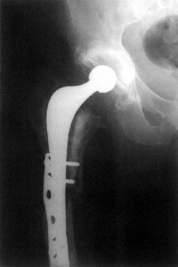
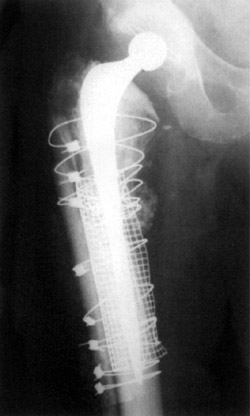
Figure 1. Third time
revision of femoral stem.
Figure 2. Allogran-N allograft impacted into femur.
Presentation
This patient was referred for a third time revision of his left THR fractured
stem and fracture shaft of femur.
Open reduction and internal fixation left femur, with strut allograft and Dall-Miles
cables. Revision total hip replacement to impaction grafted long stem Exeter.
Operation
With the patient placed on his side, and draped appropriately, the old wound
was opened and the hip approached through virgin territory posteriorly.
Dislocation was achieved easily. The acetabulum was found to be stable, and
the proximal stem was removed. The existing AO plate was removed and distal
fragment of stem retrieved, using a drill tap system. Cement was removed distally.
The large defect in the cortex was surrounded with a stainless steel mesh and
tension band wire through the linear aspira, leaving the soft tissue attachments
in place as far as possible. Four allograft struts were applied to the outside
of the femur sub-periosteally, and secured with a Dall-Miles cable system.
The femur was prepared proximally using the Exeter impaction grafting system,
Allogran-N Hydroxyapatite and two femoral heads to take a 200mm Exeter prosthesis,
with a single mix of CMW cement with Gentamycin.
Stable reduction was achieved with a neutral head. The remainder of the bone
graft/Allogran-N composite was packed around the periosteal pouch at the fracture
site posteriorly. Closure was achieved with vicryl and clips to the skin. One
drain was placed.
Surgery took 4 1/2 hours. Blood loss was 1,400mls. Antibiotic cover was Cefuroxime
1.5g, 3 doses at 6 hour intervals.
Post
Op
An X-ray was taken on the ward the morning following surgery, which confirmed
alignment. The patient was reported as being able to mobilize, and was weight
bearing to tolerance with crutches for 6 weeks, thereafter full weight bearing.
Proximal calcar was built around the mesh. The greater trochanter was held on
to the mesh with wires. A small Exeter 37.5mm offset stem prosthesis was fixed
with Gentamycin cement. On the table, neutral head height, as per the pre-operative
plan was achieved, as was a stable reduction with a long neck.
Haemostasis was secured and a thorough wash out carried out after the procedure.
A layered closure was made over two drains. Subcuticular vicryl stitches were
used as well as clips for skin closure.
Surgery took 4 hours. Antibiotic cover was Cefuroxime, following tissue sampling
and repeated 3 times at 6 hourly intervals.
Post Op
The patient's right leg seemed slightly longer. X-rays were taken on the ward,
with full weight bearing mobilization. Drains were removed at 24 hours. The
wound showing no signs of leaking.
4 days post op
The patient developed a dislocation of her right hip. This was reduced under
general anesthetic. X-rays were taken at 50° of flexion and 35° of internal
rotation, the joint was stable. With the leg fully extended, there was found
to be 2mm of shuck. The hip was stable enough for the patient to mobilize immediately.
Follow Up
2 months post op
Patient is stable, able to fully weight bear and is doing well.
6 months post op
At six months post op, the patient reported to be satisfactory, whilst still
experiencing some thigh pain attributed to a complete fracture through the pelvis.
1 year post op
Patient reported to be satisfactory, her hip pain having markedly reduced but
still persisting. There was no evidence of infection. Her hip score was measured
by self-assessment and found to be 68% (pre-op measurement was 11%).
X-rays showed substantial vertical migration of the cup measured at 6mm, this
having occurred during the first three months and then stabilizing. Stem subsidence
of 2mm was also early but did not progress.
2 years post op
Patient doing well, no further migration of the components and her hip pain
has resolved.
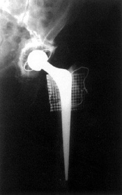
Figure 3. Post op Allogran-N impacted into the acetabulum.